


 70 / 72

with degradation of the implant via intracellular incorporation of
degraded implant material particles. For both the HA- and the HA/col-1
treated animals, remnants of the HA implant were frequently found
between ruffled borders invaginations in the cytoplasm and in
phagosomes but not in the nuclei.
Differences in the histomorphometric evaluation of new bone
formation between high-resolution peripheral quantitative computed
tomography (HR-pQCT) and
“
classical
”
histomorphometry are of
importance. The first method was unable to distinguish between
the used HA- or HA/col-1 implant from bone which made a reliable
evaluation of new bone formation within the material impossible. This
is due to the fact that the used HA in the implants closely mimics
human HA limiting the value of CT methods if such implants are used.
Therefore, the interface region was investigated by HR-pQCT and
histology with a distance of 1 mm to the initial defect. HR-pQCT
revealed the highest BV/TV ratio and smallest trabecular spacing
for the HA group and the highest connectivity density and highest
number of trabeculae for the HA/col-1 group suggesting better new
bone formation compared to the empty defect. However,
“
classical
”
histomorphometry failed to showany significant enhancement of bone
formation at the interface region between the groups.
The results of the current work are based on an established large
animal model with induction of a significant loss of the BMD by
ovariectomy and low-calcium diet in Chinese goats. The observed
reduction in BMD in this work with a decrease of BMDmeasured in the
calcanaeus of 24 ± 2% are comparable to similar studies using the same
animal model [15]. As most osteoporotic fractures affect trabecular
bone of the metapyhseal area of long bones, themetaphyseal regions of
lumbar vertebrae, of the iliac crest and of the distal femur were selected
for the current work. However, the study is limited by the small
number of animals and the low number of the specific defects.
Furthermore, only drill hole defects but no fractures were created in the
current study which limits its conclusions for the potential enhance-
ment of these two materials on fracture healing in osteoporotic
fractures [21].
Conclusion
In conclusion, both nanoparticulate HA with and without collagen
type-1 showed better new bone formation compared to untreated
drill hole defects in metaphyseal defects of the lumbar spine,
the iliac crest and in the distal femur of this osteoporotic Chinese
mountain goat model. Both materials showed good biocompatibility
without any inflammatory reaction and degradation via osteoclast-
like multinuclear cells with intracellular uptake of the material into
these cells.
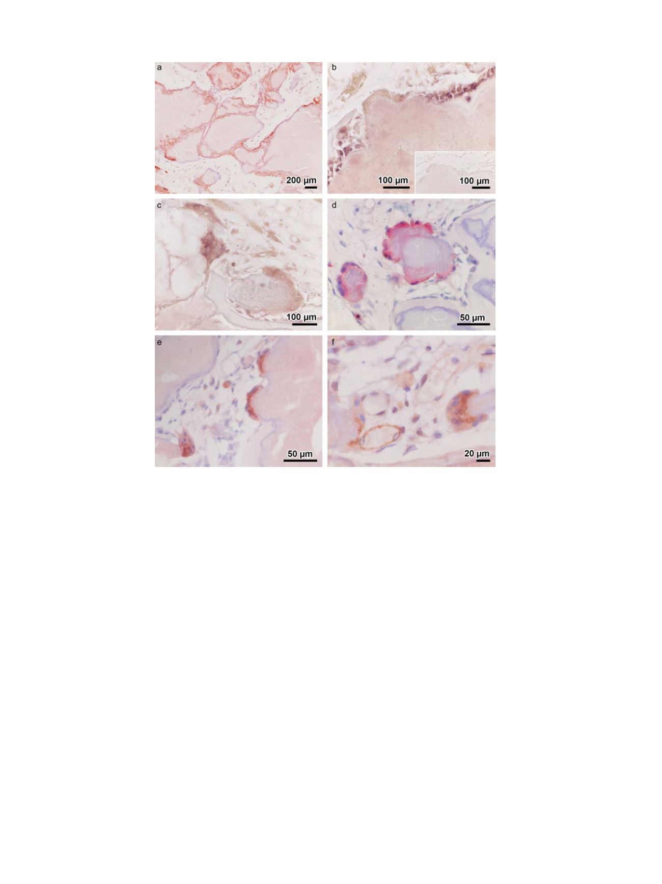
Fig. 4.
Enzyme-, immunohistochemistry, and in-situ hybridization. Osteoid surrounding single HA fragments was identified by collagen type I immunoreactivity (
a
). The
implants were covered with osteoblasts determined by connexin-43 in-situ hybridization (
b
). Inset in (b): negative control. Connexin-43 mRNA was also found in osteoclast-
like cells at the HA/col-1 implant (
c
). Osteoclast-like cells were also identified by TRAP enzyme histochemistry as shown here for the HA implant (
d
). In addition, osteoclast-like
cells were determined by CD68 immunohistochemistry as shown here at the bone-HA interface (
e
). eNOS immunoreactivity was used for identification of osteoclast-like cells as
well as newly formed blood vessels in the granulation tissue between the implant fragments (
f
).
V. Alt et al. / Injury, Int. J. Care Injured 47S2 (2016) S58
–
S65
S64