


 37 / 72

the
“
problem of osteoporosis
”
better into perspective and reduce its
relevance in the management of distal radius fractures.
Discussion
Osteoporotic fractures can occur in any bone from the dens to the
foot. In addition to the injuries already described in this article, distal
humerus fractures, insufficiency fractures of the pelvis, distal femur
fractures around the knee, ankle fractures and periprosthetic fractures
also present a great challenge to trauma surgery in the presence of
osteoporosis [37
–
43]. In this article we have discussed the most
common osteoporotic fractures, for which often different treatment
options are available and we suggest that treatment decision can be
based on bone densitymeasurement. Bone density certainly influences
the stability of surgical fracture fixation and this has been proven in
numerous experimental biomechanical studies as cited above, but
there are other reports based on clinical data that do not confirm an
unequivocal relationship between osteoporosis and failure [44,45].
Two reasons may explain this lack of clinical evidence. The studies
currently reported did not declare the analysis of an association
between failure and bone density as a primary endpoint. The available
data are the results of either retrospective analyses or secondary
endpoints of prospective studies. Furthermore, the differences in
definitions and test procedures for bone density across the various
working groups do not permit the data to be brought together for the
purpose of proper metaanalysis [44]. Another reason for the lack of
evidence for an existing relationship between BMD and failure could be
that other factors associated with the three key areas of
“
adequate
reduction, implant positioning, and implant selection
”
may have a
much greater influence on failure (Figure 4). In the
“
healthy
”
skeleton
the biological repair processes at work in fracture healing can
potentially compensate for deficits in the three key areas, but these
mechanisms are very limited in the metabolism of osteoporotic bone.
With regard to low mineralization density the published literature
offers insights into what
“
low
”
really means in the specific skeletal
regions and what the threshold value is beyond which the risk of
failure increases, whereby these BMD values that are generally derived
from CT imaging have not found application in clinical routine despite
the availability of the data. This is certainly due in part to the difficulties
surrounding QCT testing in the preoperative setting but another reason
for the absence of widespread application may be ignorance of the
data, which is often obtained and disseminated only within the
confines of academic institutions. Apart from BMD values good
experience has been gained in the intraoperative evaluation of bone
quality using the DensiProbe [46
–
48]. This instrument is a potentially
handy tool in the intraoperative decision-making process, for example,
when deciding whether cement augmentation is necessary or not.
Although the body of data for the DensiProbe is very promising, this
instrument has likewise failed to achievewidespread entry into clinical
application beyond the walls of academic institutions.
Conclusion
It can be postulated based on our clinical and scientific experience
coupled with research findings from the published literature that
fracture fixation in osteoporotic bone is less promising if the
osteosynthesis is suboptimal in an environment of weakened bone
structure due to low mineralization density. Optimization of preopera-
tive diagnostics might help to revise the treatment algorithm to take
bone density into account, thus reducing the risk of failure and, at the
same time, acquiring additional data for future reference.
Conflict of interest
The authors have no conflict of interest.
References
[1]
Dash SK, Panigrahi R, Palo N, Priyadarshi A, Biswal M. Fragility hip fractures in elderly
patients in Bhubaneswar, India (2012
–
2014): a prospective multicenter study of 1031
elderly patients. Geriatr Orthop Surg Rehabil 2015;6:11
–
5.
[2]
Konstantinidis L, Papaioannou C, Hirschmuller A, Pavlidis T, Schroeter S, Sudkamp NP,
et al. Intramedullary nailing of trochanteric fractures: central or caudal positioning of
the load carrier? A biomechanical comparative study on cadaver bones. Injury
2013;44:784
–
90.
[3]
Konstantinidis L, Papaioannou C, Blanke P, Hirschmuller A, Sudkamp NP, Helwig P. Failure
after osteosynthesis of trochanteric fractures. Where is the limit of osteoporosis?
Osteoporos Int 2013;24:2701
–
6.
[4]
Augat P, Simon U, Liedert A, Claes L. Mechanics and mechano-biology of fracture healing
in normal and osteoporotic bone. Osteoporos Int 2005;16(Suppl 2):S36
–
43.
[5]
Konstantinidis L, Papaioannou C, Mehlhorn A, Hirschmüller A, Südkamp NP, Helwig P.
Salvage procedures for trochanteric femoral fractures after internal fixation failure:
biomechanical comparison of a plate fixator and the dynamic condylar screw. Proc Inst
Mech Eng H 2011;225:710
–
7.
[6]
Mehlhorn AT, Strohm PC, Müller CA, Konstantinidis L, Schmal H, Südkamp NP. The
reversed locked internal plate fixator as an alternative internal fixation of problematic
proximal femur fractures. Z Orthop Unfall 2009;147:561
–
66.
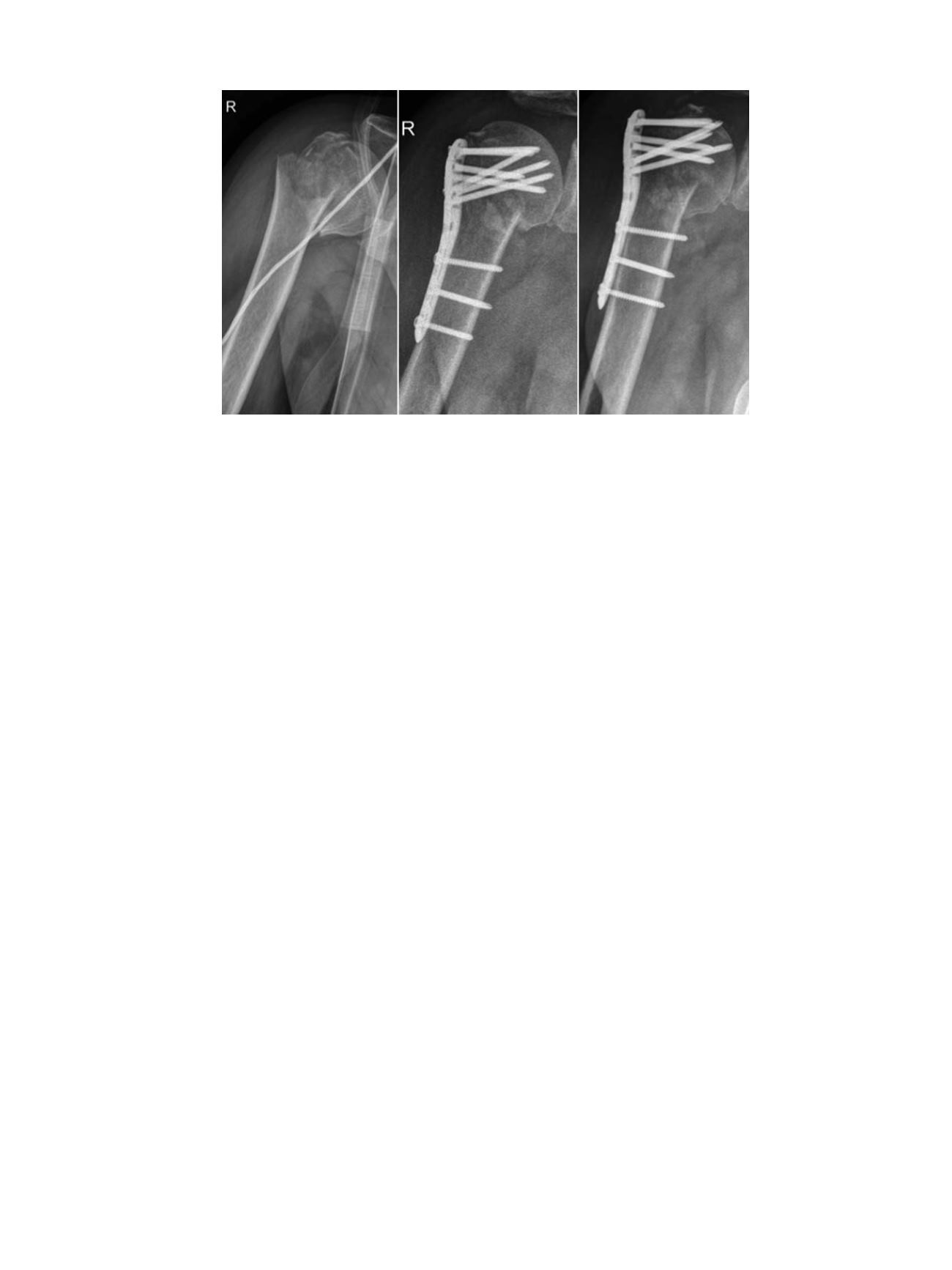
Fig. 4.
Fracture treatment (left and middle) after 4 part humeral head fracture in a 72-year-old patient. Postoperative follow up with failure at 6 weeks postoperatively (right).
Given insufficient reduction, lack of medial support and suboptimal placement of the implants failure related to bone quality was highly probable. Angular stability and bone
quality in this case could not compensate for the reduction deficits.
L. Konstantinidis et al. / Injury, Int. J. Care Injured 47S2 (2016) S27
–
S32
S31