


 34 / 72

Trochanteric femur fractures
Proximal femur fractures in the region of the trochanter are one
of the most frequent and most serious osteoporotic fractures due to
the lack of alternatives to surgical management. Surgical intervention
is unavoidable in the majority of cases; joint replacement may be
possible in principle, but will be a highly complex challenge;
osteosynthesis often remains the best therapeutic option despite the
presence of osteoporosis. Fixation failure often signals the end of
patient mobility [5,6], whereby the appropriate fixation is physiolo-
gically destined to experience relatively high loads in the region of the
hip [7]; in many cases it is not reasonable to expect partial loading
given the reduced condition of general health typical of these patients.
It is all the more important that proper consideration be given to the
afore-mentioned key concerns in fracture management
–
reduction,
appropriate choice of implant and correct implant placement. Fracture
reduction and correct implant positioning are the responsibility of
the surgeon for which he has some guiding criteria available [2].
With regard to the choice of implants, modern and clinically proven
solutions are available from most manufacturers. In particular
rotationally stable implants have clearly lowered complication rates
in osteoporotic bone in recent years [8]. Nevertheless, failures that lead
to cut out even where detailed analysis of the three key areas showed
no relevant deficits can be clinically observed. In these cases, it can be
assumed that bone quality had reached a critical threshold that limited
the efficacy of fracture fixation.
Biomechanical experiments were employed to identify a possible
threshold of bone mineral density for a reliable fixation of implants in
the proximal femur [3]. First, we tested 30 proximal femurs from
human body donors for bone mineral density (BMD) at the femoral
head using quantitative computed tomography (QCT). We selected this
region of interest because load transfer during weight bearing takes
place at the interface between the cancellous bone of the femoral
head and the femoral head screw. It is in this area that BMD is especially
important for the stable anchorage of the load carrier. After deter-
mining BMD, osteotomy was performed to simulate an unstable
trochanteric AO type 31 A2.3 fracture followed by intramedullary
nailing with insertion of the most recent generation of nails (PFNA
from Synthes, Trigen Intertan from Smith&Nephew and Targon PFT
from Aesculap). After fracture fixation cyclic dynamic loading of the
constructs was performed until failure. The primary endpoint of
the study was calculation of the relative risk of cut out in relation to the
BMD values (Figure 1). The incidence of cut-out for BMD less than
250 mg/cm
3
was 0.55 (5 of 9) and for BMD greater than 250 mg/cm
3
0.05 (1 of 21). Therefore, the risk of cut-out for BMD <250 mg/cm
3
was
almost 11 times greater than for BMD >250 mg/cm
3
. The conclusion
can be summarized as follows. There is a very high risk of implant
failure after surgical management of trochanteric fractures where BMD
is below 250 mg/cm
3
in the region of the femoral head. A threshold
value like this for bone density could be helpful, for example, when
deciding for or against cement augmentation [9] at the bone-screw
interface in the femoral head. Currently, there are no definitive
decision-making criteria for implant augmentation [10] and wide-
spread application of augmentation to all trochanteric fractures would
not be advisable because of the associated complication risks as well
as for socio-economic reasons. On the other hand, determining bone
density in the region of the femoral head is not easy logistically. In
principle, it is not very difficult to perform QCT, however, some
institutions do not have the necessary infrastructure and the software
of the CT manufacturers is often not sophisticated enough for this
special application. Despite these constraints and based on our own
experimental findings and data from the literature [11], a BMD
threshold of 250 mg/cm
3
appears to be a clinically relevant values for
the prediction of stability of intramedullary osteosynthesis of proximal
femur fractures.
Medial femoral neck fractures
Medial femoral neck fractures occur at an incidence similar to that
of trochanteric fractures and are likewise a typical osteoporotic fracture
type. In practice, treatment depends on the classification of the
fracture, whereby international and national directives for fracture
management do not provide practical guidelines and leave the surgeon
great freedom to make treatment decisions. Nevertheless, it can be
broadly stated that stable femoral neck fractures should be treated by
osteosynthesis [12] and unstable fractures by joint replacement [13].
Osteosynthesis of stable fractures is not susceptible to any relevant
mechanical failures in the sense of cut-out or fracture dislocation [12],
provided that the classification of the fracture as stable or unstable is
correct. The risk of complications does however increase for unstable
fractures, but since joint replacement surgery is available for unstable
femoral neck fractures and produces excellent functional results [13],
alloplastic treatment is a viable option for unstable fractures with
osteoporosis. Given this situation, we see no indication for further
differentiation or analysis of bone quality in relation to fracture fixation
for medial femoral neck fractures.
Vertebral body fractures
Pathological vertebral body fractures without relevant trauma or
after low energy trauma represent a major challenge in the context
of osteoporosis. In many cases, non-surgical treatment is extremely
promising and offers satisfactory functional outcomes [14]. However,
in some cases surgery is indicated, either to alleviate pain or to reverse
some marked deformity such as spinal canal stenosis, which is
associated with pain and neurological deficits. This section is con-
cerned with the challenges of screw fixation in osteoporotic vertebral
bodies, but not with vertebroplasty or kyphoplasty, which represent
more or less invasive approaches to pain therapy with low levels of
evidence to date [15].
Typical failures of dorsal instrumentation are cut out and also pull-
out of the pedicle screws [16]. In contrast to trochanteric fractures it
is bone quality that is more frequently responsible for failure rather
than reduction or precise screw placement, which is generally exactly
transpedicular because of the anatomical features of the region.
Various technical methods are available if the treatment of choice for
osteoporotic vertebral fractures is dorsal instrumentation: Simple,
bisegmental dorsal bridging of the fractured vertebra is the
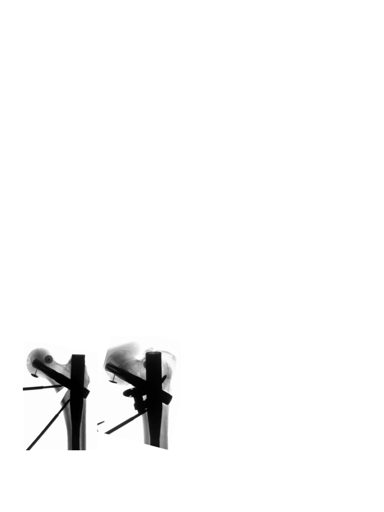
Fig. 1.
Radiological images of a construct incorporating proximal femoral nail osteo-
synthesis (PFNA, DePuy-Synthes) before loading (left) and after 10,000 load cycles
(right) at 2100N. It shows cut out typical of a clinical complication involving mediali-
zation of the PFNA blade, varus dislocation and collapse of the osteotomy gap, which
corresponds to comminution in the clinical environment.
L. Konstantinidis et al. / Injury, Int. J. Care Injured 47S2 (2016) S27
–
S32
S28