


 20 / 72

evidence exists that this heterogeneity has important biomechanical
consequences, but further work is required to establish mechanisms
and clinical implementation of these insights.
Tissue heterogeneity
Changes in tissue composition and mechanical properties at the
material/tissue level (lamellae, individual trabeculae) likely contribute
to fracture risk, but up until recently these changes have been less well
understood. A number of studies have sought to address this, using a
combination of mechanical testing (nano-indentation, micro-mech-
anical testing) [56
–
60] and compositional analyses at the tissue level
[57
–
59,61
–
64], and their findings regarding changes in tissue
properties and composition during osteoporosis are conflicting. It
has been reported for example that trabecular bone tissue from the
proximal femur of ovariectomized sheep (12months post-surgery) had
a lower tissue modulus, as measured by nano-indentation, compared
to age matched controls [56,57]. These changes were associated with a
decrease in mineral content in the osteoporotic trabecular bone tissue
[57,62]. Interestingly, the differences were not maintained 31 months
post-surgery [57]. In contrast, micro-tensile testing showed that the
stiffness and strength of ovariectomized rat trabeculae was increased
by 40
–
90% by 54 weeks post-ovariectomy [58,59]. These increases
were associated with a significant increase (11%) in the mineral content
of these trabeculae
,
although overall bone mineral density and mass
were reduced [58,59]. It has also been reported that increased calcium
content and stiffness occur within individual trabeculae from human
osteoporotic bone [64,65].
Variations in experimental methods, animal model or the anatom-
ical location from which bone was chosen for analysis might explain
the discrepancies between previous studies. For example decreased
trabecular stiffness was reported based on nanoindentation of
trabeculae from the anteromedial region of the proximal femur of
the ovariectomized sheep [56,57], whereas increased trabecular
stiffness was based on micro-tensile testing of trabeculae from a
region below the growth plate of the tibia of ovariectomized rat bones
[58,59]. Nanoindentation characterises the mechanical properties
(elastic modulus, hardness) of nanometer areas of bone tissue
(typically within individual lamellae), whereas micro-tensile testing
assesses the mechanical behaviour of entire trabeculae. Therefore, to
understand these discrepancies further a recent study sought to
distinguish (1) the spatial distribution of mineral within different
lamellae across individual trabeculae and (2) the variation in trabecular
mineralisation in different anatomical regions of the proximal femur
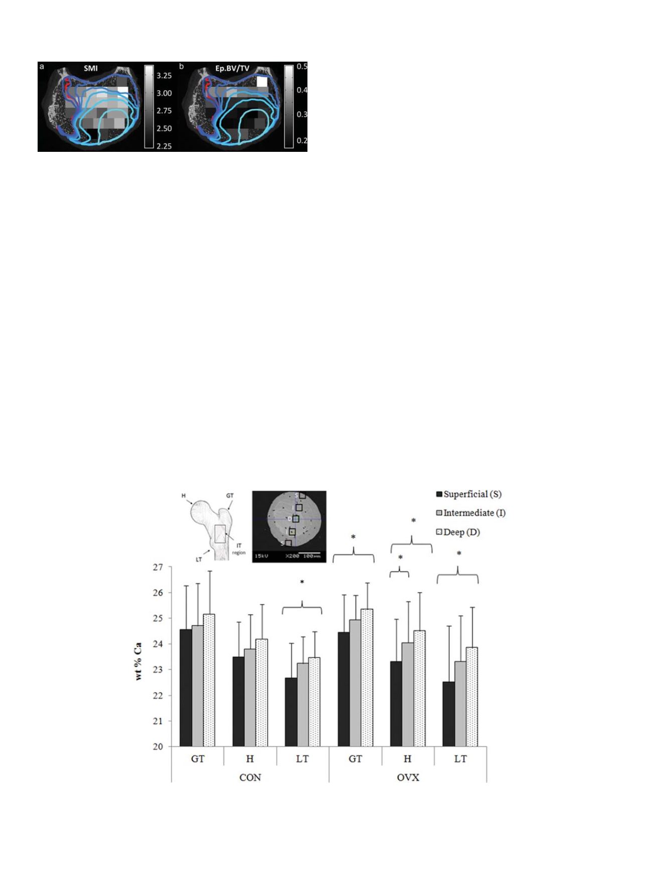
following the onset of estrogen deficiency [66]. Mineral content (wt%
Ca) was determined using a quantitative backscattered scanning
electron microscopy approach, for individual trabeculae harvested
from the proximal femur of ovariectomized sheep (12 months post-
OVX) and age-matched controls. It was found that the difference in
mineralization between the superficial and deep lamellae of trabeculae
was more pronounced in ovariectomized sheep (Figure 5), represent-
ing an increase in mineral heterogeneity of approximately 13%,
compared to trabeculae from aged matched controls [66]. Moreover
Fig. 4. Bone heterogeneity and vertebral endplate collapse
Regions of endplate col-
lapse (outlined in blue and red) and distribution of structure model index (SMI) in
the trabecular bone directly underlying the endplate (grayscale): The lightest blue
outline corresponds to the loading increment at which endplate collapse clearly
initiated. The boundaries at subsequent loading increments are represented with
progressively darker shades of blue. The red outline corresponds to the region of
endplate collapse that remained after loading was complete and all load was
removed. Modified from Jackman et al. [52].
Fig. 5. Trabecular mineralization in estrogen deficiency
Spatial distribution of calcium (wt% Ca) between superficial, intermediate, and deep lamellae in the greater trochanter
(GT), head (H) and lesser trochanter (LT) regions of the proximal femur from 12 month ovariectomized sheep (OVX) and aged matched controls (CON). * indicates statistical sig-
nificance between trabecular regions indicated by brackets (
p
≤
0.02). Figure adapted and data from [64].
G. Osterhoff et al. / Injury, Int. J. Care Injured 47S2 (2016) S11
–
S20
S14